
Light microscopy allows discovering the microcosmos that surrounds us with our own eyes using the visible light. By creating a spacial images of a given specimen, microscopy is a formidable tool to explore tiny structures and explore the spacial organisation of small organisms, tissues and cells.
The respective domains (physics, chemistry and biology) that enabled the development of microscopy hardware and sample preparation methods are intertwined and inspired each other throughout recent history of light microscopy. Our Bioimage Core Facility can also be understood as an intersection of these domains in the context of light-microscopy.
-
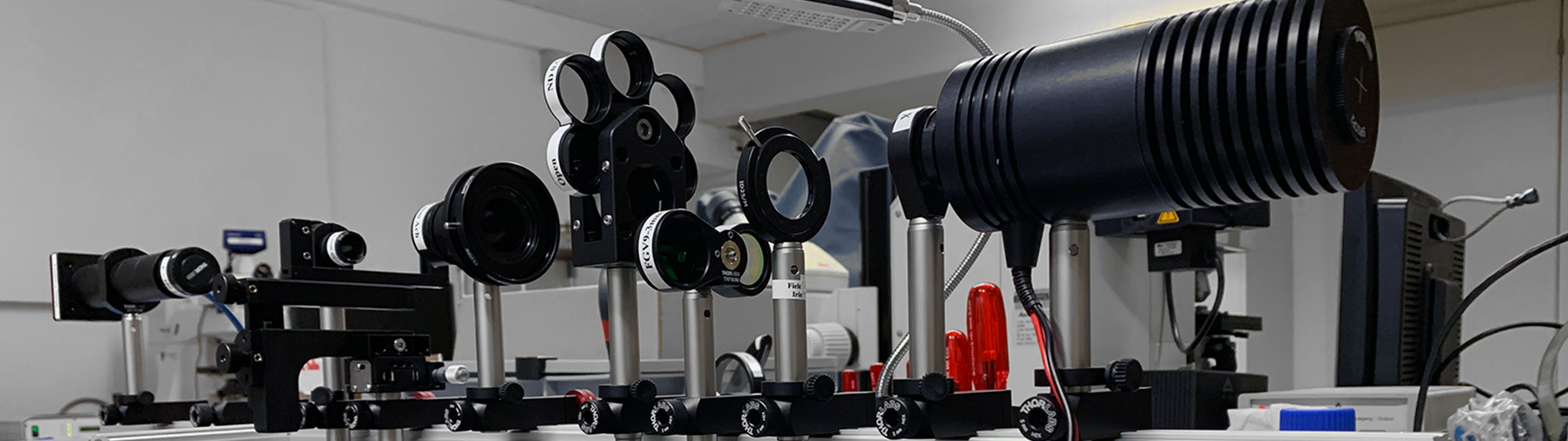
Key hardware components
- Objectives - Advances in glass manufacturing and optics made complex assemblies of lenses capable of correcting spherical and chromatic aberrations.
- Light sources - LASER and LED allow for homogeneous and precise sample illumination.
- High-speed scanning galvo mirror.
- Filters and dichroic mirrors - Modern thin-film manufacturing provides the tool to accurately control the light wavelength in the optical path.
- Point detectors: photomultiplier tubes (PMTs) and avalanche photodiodes (APDs).
- Digital cameras: EMCCD, sCMOS, and hybrid detectors enable photon counting (high sensitivity) at ever faster frame rates.
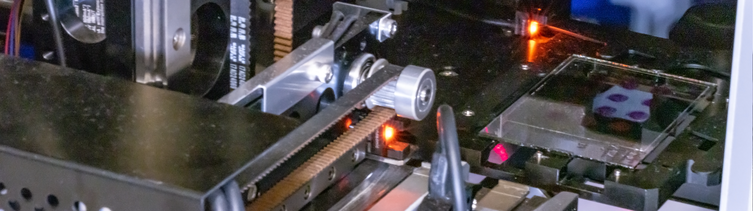
- Precision step motors for fast, coordinated sample positioning.
- Modern computers - Data transfer bandwidth and processing power are a necessity for digital cameras. Furthermore, they enable post-acquisition processing of the data (image restoration, 3D reconstruction, quantification).
-
Sample preparation milestones
- Microtomes - Studying serial thin sections of a given specimen.
- Biomarkers - Colour and fluorescent dyes enable contrast enhancement.
- Antibody staining - Colouring specific structures in tissue and organisms.
- Fluorescent proteins - Render proteins visible by attaching a fluorescent sequence that is (re)produced by the organism itself.
- Fluorescent biosensors - Making functional aspects (in contrast to mere structure) or molecules visible.
- Tissue clearing - Allows light microscopy to be applied to thicker (bigger) samples - An alternative to serial sectioning.
- Spontaneously blinking and transiently binding fluorophores.
Microscopy Techniques
The above mentioned knowledge and technologie lead to the development and implementation of numerous microscopy techniques and different setups. In the table bellow you find a list of well established microcopy techniques. Not all of these techniques are available through the core facility or a particular laboratory at our University. However, the microscopy core facilities in Switzerland are well networked. If a particular instrument is not available on campus, our staff will direct you to the closest core facility that has the instrument in question available.
| Technique | Description | Sample |
Lateral, axial - res. |
Availability |
|---|---|---|---|---|
| Bright-field1 | Transmitted light | Thin, translucent | 300nm, - |
|
| Epi-Fluorescence | Epi-illumination | Fluorescent labels | 300nm, 1um |
|
| FRET2 | Molecular interaction (proximity) | Fluorophore FRET-pair | 1-10nm, - |
|
| FRAP3 | Protein production and diffusion | Fluorescent protein | - | |
| FLIM/TCSPC9 | Fluorescent life-time measurement | Any fluorescence | - | |
| QPI/(DHM) | Label-free, 3D | Thin, translucent | 200nm, 400nm |
|
| TDM/(DHM) | Label-free, 3D | Thin, translucent |
150nm, 180nm |
|
| TIRF/(SNOM)4 | Evanescent excitation light | Fluorophore near coverslip | 230nm, 100nm |
|
| Laser scanning confocal | High contrast, up to ~300um deep. | Fluorescent labels | 200nm, 500nm |
|
| Array detector (Airyscan, NSPARC) | Fluorescent labels |
100nm, 200nm |
||
| Spinning disk confocal | Fast, high contrast, up to ~150um deep. | Fluorecent labels | 200nm, 500nm |
|
| Multi-photon | Confocal for deep tissue imaging | Special fluorophores | 300nm, 1um |
|
| SIM | Fast super resolution | Fluorecent labels | 100nm, 300nm |
|
| STED | 3D super resolution | Special fluorophores | 50nm, 100nm |
|
| Light sheet | Fast, photon-economic 3D imaging. | Sample holder compatible |
500nm, 2um ?? |
|
| Single molecule detection |
PALM7, STORM8 |
Photoactivatable or blinking fluorophores | 10–100nm, ?? |
|
|
1: Incl. dark-field, phase contrast, differential interference contrast (DIC) , polarised light 2: Förster resonance energy transfer 3: Fluorescence recovery after photobleaching 10: Quantitative phase imaging / digital holographic microscopy 11: Tomographic diffraction microscopy / digital holographic microscopy |
: Available in core facilities : Existing in research labs : Not available on campus |
|||