Dynamic light scattering (DLS) is an analytical technique used to determine the size distribution of small particles, such as nanoparticles, in suspension by measuring fluctuations in the intensity of scattered light caused by the Brownian motion of the particles.
When a laser beam passes through a colloidal sample, the scattered light's intensity varies over time, and this variation is analyzed to calculate the particles' hydrodynamic diameter. DLS is widely used because it is straightforward and rapid.
Developed in the mid-20th century as laser and photodetector technology advanced, DLS has become a standard tool for characterizing nanoparticles, polymers, proteins, and colloids in various scientific fields. Its applications span materials science, nanotechnology, pharmaceuticals, and biopharmaceutical development, where it is used to assess particle size, aggregation, and colloidal stability in formulations and nanomaterial research. 3D dynamic light scattering (3D-DLS) is an advanced form of DLS that uses multiple detection angles or cross-correlation schemes to improve the accuracy and reliability of particle size and dynamic measurements, especially in complex or turbid samples. By capturing scattered light from several directions and employing cross-correlation, 3D-DLS can better distinguish true particle motion from artifacts or multiple scattering, making it particularly valuable for challenging samples such as concentrated protein solutions, nanoparticle suspensions, or biofluids
Directly to the Instrument:
-
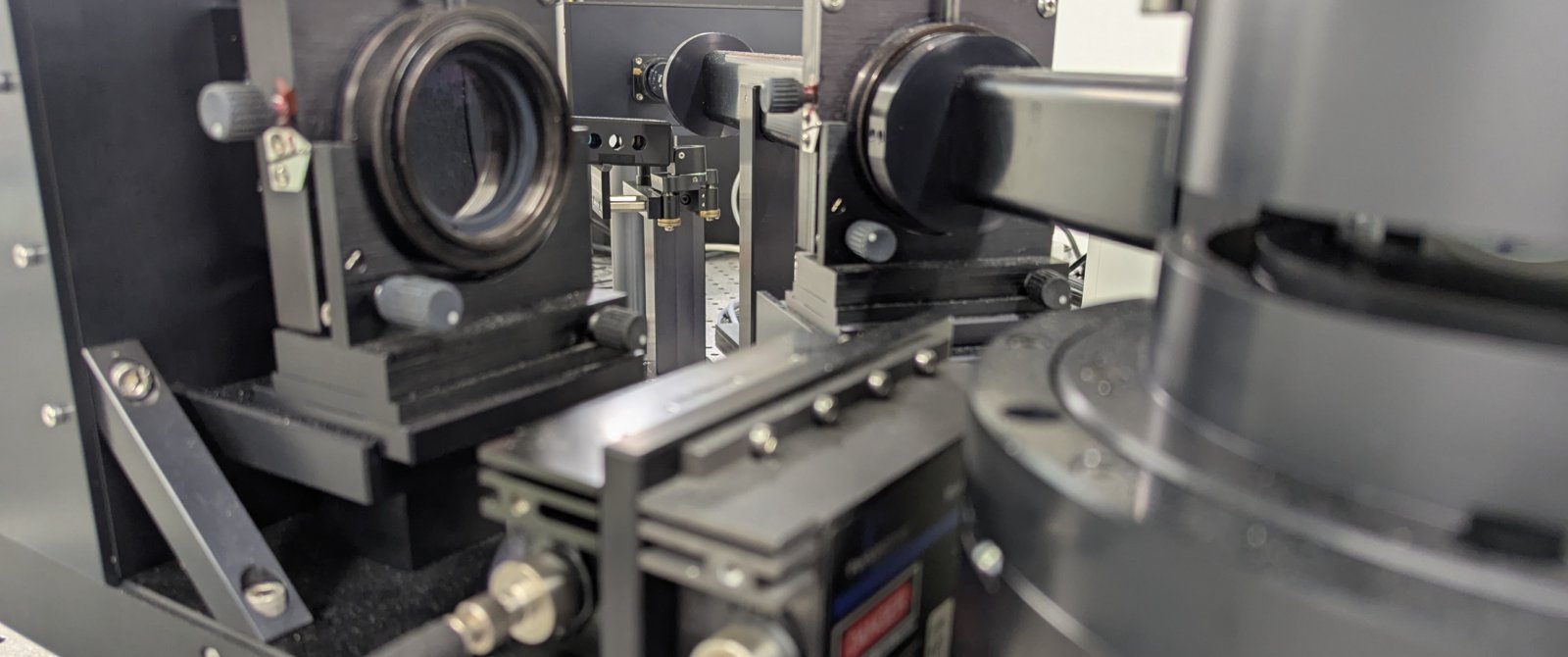
Key hardware components
- Laser source: Provides a coherent, monochromatic light beam that illuminates the sample.
- Sample holder (cuvette or capillary): Contains the particle suspension or solution to be analyzed.
- Photodetector (such as a photomultiplier tube or silicon photomultiplier): Detects the intensity of light scattered by the particles in the sample.
- Correlator or digital oscilloscope: Processes the fluctuating scattered light intensity data to analyze particle motion and calculate size distribution.
- Computer and control electronics: Collect, process, and display the data for interpretation and analysis.
-
Sample preparation milestones
Proper sample preparation is crucial for reliable dynamic light scattering measurements, as DLS is highly sensitive to contaminants, aggregates, and sample heterogeneity.
Key Considerations
- Clean labware and filtered solvents are essential to minimize contamination.
- Automation and standardization of sample preparation can improve reproducibility and reliability, especially for nanomaterial analysis.
- For oxidation-prone or sensitive nanoparticles, preparation under inert atmosphere or with stabilizing agents may be necessary to preserve sample integrity
Sample preparation
- Dispersion: The sample (nanoparticles, proteins, polymers, etc.) is dispersed in a suitable solvent, often water or another medium compatible with the particles and the measurement objectives.
- Filtration or Centrifugation: Samples are typically filtered (using 0.2–0.45 µm filters) or centrifuged to remove dust, large aggregates, or debris, which can skew size measurements.
- Concentration Adjustment: The particle concentration is optimized to ensure sufficient scattering without causing multiple scattering effects; too high or too low concentrations can both lead to inaccurate results.
- Solvent Selection: The choice of solvent or dispersion medium (e.g., water, ethanol, glycerin) affects viscosity and refractive index, which are critical parameters for accurate size determination
Sample Preparation for Different DLS Modes
- Standard DLS (Particle Size Analysis): Focuses on obtaining a stable, monodisperse suspension. Removal of aggregates and dust is especially important, and the sample should be homogeneous and well-dispersed.
- Electrophoretic Light Scattering (ELS, for Zeta Potential): In addition to the above, the ionic strength and pH of the dispersion medium may be adjusted to mimic physiological or experimental conditions, as these factors influence particle surface charge and mobility.
- Polydisperse or Complex Samples: For heterogeneous or polydisperse mixtures (e.g., industrial nanopowders), more elaborate preparation such as embedding, cross-sectioning, or the use of surfactants to prevent aggregation may be required to achieve representative and reproducible measurements
Sample Preparation for 3D-DLS
- Tracer Particles: For microrheology applications, carefully selected surface-functionalized tracer particles are added to the sample to probe its properties without introducing artifacts. The optimal size and concentration of these tracers are chosen based on the sample’s characteristics to avoid aggregation or depletion effects.
- Dilution and Homogenization: Samples, especially biological fluids or nanoparticle suspensions, may require dilution to minimize multiple scattering and ensure uniform particle distribution. Homogenization ensures that particles are evenly dispersed.
- Avoiding Contaminants: All containers and solutions should be clean and free from dust or aggregates, as contaminants can skew results.
- Parameter Optimization: The amount of sample, tracer concentration, and measurement parameters are optimized to balance sensitivity and minimize artifacts, especially when dealing with high concentrations or complex mixtures
Light scattering Techniques
The above-mentioned knowledge and technology led to the development and implementation of numerous microscopy techniques and different setups. In the table below, you will find a list of well-established microscopy techniques. Not all of these techniques are available through the core facility or a particular laboratory at our university. However, the microscopy core facilities in Switzerland are well networked. If a particular instrument is not available on campus, our staff will direct you to the closest core facility that has the instrument in question available.
| Mode/Modality | Description & Principle | Applications | Possible Sample Types |
|---|---|---|---|
| Standard DLS (Single-Angle DLS) | Measures time-dependent fluctuations in scattered light at a single angle to determine particle size distribution based on Brownian motion. | Nanoparticle sizing, colloid stability, protein aggregation, polymer characterization | Nanoparticles, proteins, polymers, colloidal suspensions, exosomes |
| Multi-Angle DLS (MADLS) | Collects scattering data at multiple angles to improve accuracy and resolve polydispersity and shape effects. | Detailed size distribution, shape analysis, detection of aggregates | Nanoparticles, polydisperse colloids, protein complexes |
| 3D Cross-Correlation DLS (3D-DLS) | Uses two or more detectors at different angles to suppress multiple scattering and enhance measurement in turbid or concentrated samples. | Nanoparticle-protein interactions, analysis in complex biofluids, concentrated suspensions | Gold nanoparticles, proteins, serum, dense colloids |
| MicroDLS (DLS Microscopy) | Integrates DLS with optical microscopy to enable local, high-resolution DLS measurements in small sample volumes or heterogeneous samples. | Analysis of highly concentrated or heterogeneous samples, time-evolving systems | Nanoparticles in microfluidic devices, concentrated suspensions, biological samples |
| Polarization-Resolved DLS | Adjusts polarization of detected/scattered light to minimize artifacts and enhance sensitivity to specific dynamics (e.g., flow, orientation). | Blood flow imaging, tissue perfusion, microcirculation studies | Biological tissues, blood, brain cortex |
| Laser Doppler Flowmetry (LDF) | Measures frequency shifts in scattered light due to moving particles (Doppler effect), often used for flow measurements. | Blood flow monitoring, microcirculation assessment | Biological tissues, skin, organ perfusion |
| Diffusing Wave Spectroscopy (DWS) | Extension of DLS for highly scattering (opaque) samples, analyzing deep tissue or dense suspensions. | Deep tissue dynamics, viscoelasticity of dense colloids, food, and biomedical samples | Dense colloids, food emulsions, biological tissues |