Atomic force microscopy is a high-resolution imaging technique that uses a sharp probe attached to a flexible cantilever to scan the surface of a sample, measuring the forces between the tip and the sample to generate detailed topographical maps at the nanometer or even atomic scale.
Invented in 1986 as an extension of the scanning tunneling microscope, AFM opened new possibilities for imaging and manipulating surfaces that do not conduct electricity. Today, AFM is widely applied in fields such as biology, materials science, physics, and nanotechnology, enabling researchers to study the surface structure, mechanical properties, and molecular interactions of a vast range of materials—including living cells, polymers, semiconductors, and even plant cell walls.
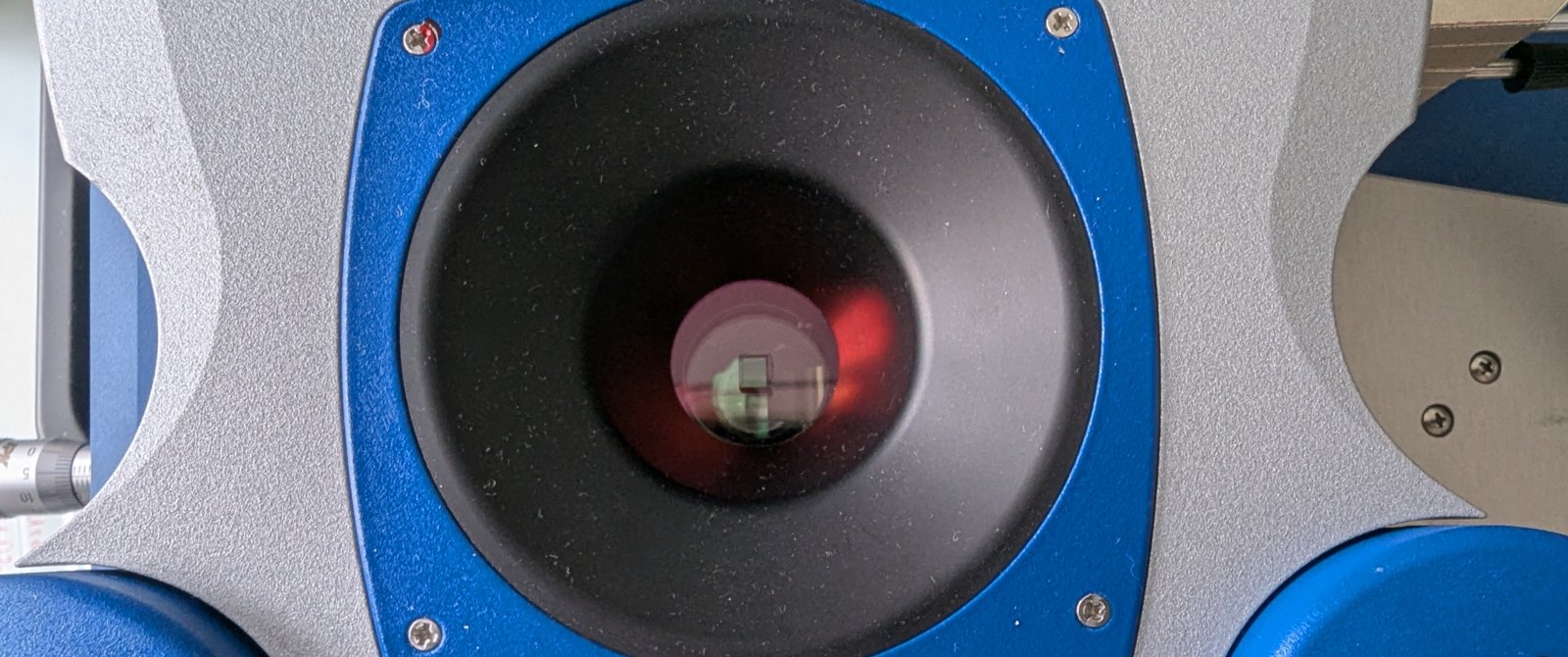
Directly to the instrument:
-
Key hardware components
-
Cantilever and sharp probe (tip): The cantilever holds the nanometer-scale tip that physically interacts with the sample surface, detecting forces at the atomic level.
-
Scanner (usually piezoelectric): Precisely moves either the sample or the cantilever in the x, y, and z directions to scan the surface at nanometer resolution.
-
Deflection detection system (often optical beam deflection): Measures the deflection of the cantilever as it responds to surface features, typically using a laser beam reflected onto a position-sensitive photodetector.
-
Sample stage: Holds and positions the sample under the probe for scanning.
-
Feedback and control electronics: Maintain constant force or height between the tip and the sample, and process signals for image formation.
-
Vibration isolation system: Minimizes external vibrations to ensure high-resolution imaging.
-
Computer and software: Control the instrument, acquire data, and reconstruct topographical images.
-
-
Sample preparation milestones
General Principles
-
The sample surface must be clean, flat, and securely immobilized on a substrate to prevent movement during scanning.
-
For many materials, straightforward cutting, grinding, or embedding and sectioning can be used, but care must be taken to avoid introducing surface roughness or smeared coatings, especially for layered or coated materials.
Contact Mode
-
Samples should be firmly attached to a flat substrate (such as mica, glass, or silicon wafer) to minimize drift and vibration.
-
For biological samples or soft materials, fixation (e.g., with paraformaldehyde) or gentle drying may be used to preserve structure and prevent deformation.
Tapping (Intermittent Contact) Mode
-
This mode is more forgiving for soft or loosely attached samples, as it reduces lateral forces.
-
Biological samples, polymers, and delicate nanostructures often benefit from this mode, with preparation focusing on gentle immobilization and, if needed, cryofixation to maintain native morphology.
Non-Contact Mode
-
Requires extremely clean and atomically flat surfaces, as the tip never touches the sample.
-
Often used for imaging hard, clean surfaces such as crystals or semiconductor wafers.
Specialized Preparations
-
For cross-sectional imaging (e.g., in wood-based materials), embedding and precise sectioning are necessary to avoid surface artifacts and to reveal internal structures.
-
For single cells or tissues, fixation (chemical or cryogenic) is often used to preserve morphology and elemental composition, with cryofixation providing the closest-to-native state for sensitive biological samples.
-
For soft or heterogeneous materials, methods may include gentle drying, critical point drying, or supporting the sample on a compliant substrate to avoid deformation.
-
-
Sample specific considerations
Polymeric Films
- Polymeric films are typically prepared by spin-coating or drop-casting a dilute polymer solution onto a flat substrate such as mica, silicon wafer, or glass. This ensures the formation of a thin, uniform, and smooth film suitable for AFM imaging.
- The substrate must be clean to avoid contamination and artifacts. Sometimes, films are annealed or solvent-vapor treated to improve surface uniformity and remove residual solvent.
- For soft or sticky polymers, care is taken to minimize deformation by using tapping mode rather than contact mode during imaging.
Nanoparticles
- For nanoparticles, a common method is to deposit a small volume of nanoparticle suspension onto a clean, flat substrate and allow it to air dry (drop-casting). However, this can lead to aggregation and uneven distribution, so advanced techniques such as ultracentrifugation or substrate functionalization are used to improve particle attachment and uniformity.
- Surface functionalization of the substrate or addition of cations to the suspension can enhance nanoparticle adhesion and distribution, minimizing aggregation and improving representativeness for quantitative analysis.
- Special attention is needed for soft nanoparticles, such as polystyrene latex, as they can deform during both preparation and imaging. This deformation can affect height measurements and must be considered in data interpretation.
Biological tissue
Fixation: Chemical fixation using agents like paraformaldehyde is common, as it preserves cell morphology and provides robust, reproducible results. Paraformaldehyde at 3.7% is widely used and balances preservation with minimal artifact introduction. Organic solvent fixation (e.g., ethanol, methanol) can cause significant dehydration and elemental redistribution, which may not be ideal for all studies.
Immobilization: Cells must be securely attached to a substrate (such as glass, mica, or functionalized surfaces) to prevent movement during scanning. Techniques include gentle drying, use of adhesive coatings (e.g., poly-L-lysine), or encapsulation in hydrogels to maintain viability and positioning.
Specialized Techniques: Advanced preparations, such as unroofing (removal of the apical membrane by sonication), enable direct AFM access to intracellular structures like the cytoskeleton for high-resolution imaging.
Microscopy Techniques
The above-mentioned knowledge and technology led to the development and implementation of numerous microscopy techniques and different setups. In the table below, you will find a list of well-established microscopy techniques. Not all of these techniques are available through the core facility or a particular laboratory at our university. However, the microscopy core facilities in Switzerland are well networked. If a particular instrument is not available on campus, our staff will direct you to the closest core facility that has the instrument in question available.
| Mode/Modality | Description/Principle | Applications | Possible Sample Types |
|---|---|---|---|
| Contact Mode | Tip remains in continuous contact with the surface | Topography, roughness, friction, wear studies | Hard materials, polymers, fixed cells, thin films |
| Tapping Mode (Intermittent Contact) | Tip oscillates and intermittently touches the surface | Soft/fragile sample imaging, biological structures, polymers | Live/fixed cells, soft tissues, gels, polymers |
| Non-Contact Mode | Tip oscillates near the surface, never making contact | Imaging delicate, loosely bound, or contaminated surfaces | Clean crystals, thin films, molecular layers |
| Force Spectroscopy | Measures force-distance curves between tip and sample | Mechanical properties (stiffness, adhesion), molecular binding | Proteins, DNA, cells, polymers, nanomaterials |
| High-Speed AFM | Rapid scanning for real-time imaging | Dynamic biological processes, protein conformational changes | Proteins, membranes, live cells |
| Conductive AFM (C-AFM) | Measures electrical current through tip-sample contact | Local conductivity, electronic device characterization | Semiconductors, nanowires, organic electronics |
| Magnetic Force Microscopy (MFM) | Detects magnetic interactions between tip and sample | Magnetic domain imaging, data storage research | Magnetic materials, thin films, nanoparticles |
| Electrostatic Force Microscopy (EFM) | Maps electrostatic forces | Surface potential, charge distribution | Electronic devices, polymers, biomolecules |
| Kelvin Probe Force Microscopy (KPFM) | Measures work function and surface potential | Semiconductor research, corrosion studies | Metals, semiconductors, organic films |
| Lateral Force Microscopy (LFM) | Measures frictional forces as tip scans sideways | Friction, wear, lubrication studies | Polymers, composites, coatings, biological films |
| Nanoindentation | Tip indents surface to measure hardness and elasticity | Mechanical property mapping | Polymers, biomaterials, thin films, tissues |
| Chemical Force Microscopy (CFM) | Functionalized tips probe specific chemical interactions | Mapping chemical groups, biomolecular recognition | Proteins, DNA, functionalized surfaces |
| Photothermal AFM | Combines AFM with photothermal excitation | Thermal property mapping, photothermal response studies | Polymers, nanocomposites, biological samples |
| AFM-IR (Infrared AFM) | Combines AFM with IR spectroscopy | Nanoscale chemical identification, polymer blends | Polymers, biomolecules, thin films |
| Scanning Thermal Microscopy (SThM) | Measures local temperature and thermal conductivity | Thermal property mapping, electronics research | Microelectronics, polymers, composites |
| AFM-Based Nanolithography/Manipulation | Uses tip to modify or move material at nanoscale | Nanopatterning, molecular manipulation | Polymers, nanomaterials, DNA, proteins |