Fabienne Marie Perren
Maître-assistant·e
Brain circulation has unique structural and functional properties.
The LUNIC Laboratory projects using and developing ultrafast ultrasound localization microscopy may facilitate the understanding of brain hemodynamics and of how vascular abnormalities in the brain are related to neurological pathologies.
The Laboratory of ultrafast Ultrasound NeuroImaging in Clinics (LUNIC) is an interdisciplinary team of neurologists, physicists, psychologists and post-doctoral students directed by PD Dr. Perren.
Our research group works between the Section of Medicine Neurology Unit of the University and Hospital of Fribourg, the Geneva Neurocenter, the CRNS and the EPFL-Campus Biotech.
We welcome Bachelor and Master medical students who are interested in cerebral hemodynamics and neurosciences.
LUNIC Laboratory projects, based on ultrafast ultrasound, a revolutionary noninvasive imaging tool, developed in humans in our group, are dealing with its methodological developments in neurology and, in particular, in cerebrovascular diseases. By using plane-wave emissions rather than line-by-line pulse echo, frame rates can be increased from 50 to 20 000 Hz giving access to rapid phenomena. Our projects are intended to assess biomechanical properties of the cerebral vessels in various pathologies (aneurysm, vasculitis, atheromatosis, dementia), microcirculation (functional imaging), cerebrovascular blood flow and perfusion (modification, regulation) in ischemia (stroke, vasospasm) and in intracranial hyper- or hypotension.
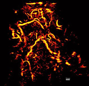
ULM, by localizing intravenously injected microbubbles, has achieved transcranial imaging of cerebrovascular flow, up to micrometer scales. This methods allows to reconstruct the finest vessels.
We are working on the optimization of the localization precision, microbubble separation, acquisition time, tracking, and velocimetry to improve the capacity of ULM to detect and distinguish vessels much smaller than the wavelength.
We also work on ULM application in vivo in clinics in the brain. Indeed, ULM is bound to improve drastically
our vision of the microvasculature. We think that it could revolutionize the comprehension, detection and diagnosis of several cerebral and cerebrovascular pathologies.
Dr Perren is teaching a part of the Clinical Competences (CC) in Neurology during the third year of medical school at the University of Fribourg.
The teaching is organized as follow:
Clinical competences (8h with groups of 5 students)