Services
The BSI Platform provides comprehensive theoretical teaching, hands-on training, support and counseling for electrical neuroimaging (EEG), non-invasive brain stimulation, magnetic resonnance imaging and neuropsychology.
-
Electrical neuroimaging
We have expertise in using high-density electroencephalography (EEG) as a neuroimaging technique with millisecond temporal resolution. We analyze ERP using Electrical Neuroimaging methods based on a step-wise procedure that allows the statistical assessment of (1) ERP morphology and amplitude, (2) global electric field power, (3) topographic stability of and changes in the electric field configuration, and (4) intracranial distributed linear source estimations (see e.g. Brunet, Murray & Michel, 2010). These analyses provide data-driven, statistically-based neurophysiological interpretation of scalp-recorded ERPs. Specifically, these analysis methods utilize the data from all channels, thereby avoiding a major source of experimenter bias, notably including (arbitrary) choice of the reference and of period and electrode of interest. In addition to statistically identifying the timing of response modulations (as in standard waveform analysis procedures), these methods differentiate effects due to (a) strength modulations of statistically indistinguishable generator configurations, (b) latency shifts across conditions, (c) topographic differences (and therefore changes in the active brain sources), or (d) any combination of these neurophysiologic phenomena, since these analyses are independent. In parallel, we have expertise in methods for the solution of the so-called inverse problem, which in the case of ERPs refers to the estimation of the activity of intracranial sources based on scalp-recorded data with biophysically-based a priori constraints to obtain a unique source estimate. These methods allow us not only to estimate brain activity for each voxel of our solution space at each moment in time, but also to perform statistical analyses with these estimations across experimental conditions (e.g. Manuel et al., 2010). The principal advantage of electrical neuroimaging techniques is their capacity to address when during a task different mental processes are engaged, and by extension in what sequence(s) different configurations of active areas are involved. Such information is critical for addressing issues of sequential versus parallel activity, feedback vs. feedforward processing, etc. at the core of our projects In close collaboration with the Center for Biomedical Imaging of the CHUV, we also use single-trial classification method based on topographic analysis comprising two steps (e.g. Bernasconi et al., 2011; Tzovara et al., 2012). First, a mixture of Gaussians model identifies representative topographies irrespective of their timing and trials; second, posterior probabilities for each Gaussian provide statistical inference on the structure of these topographies across trials, time, and experimental conditions. These methods intend detailing interdependencies between single-trial responses and performance. In clinical research, it gives the possibility to statistically evaluate single subject data, an essential tool for analyzing patients with specific deficits and that cannot be considered part of a group and/or if not enough patient can be found to constitute homogeneous clinical groups.

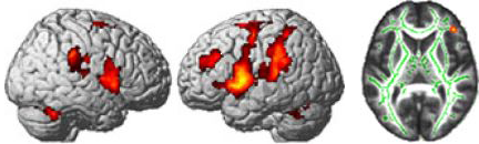
Up-left: EEG cap with color-coded instantaneous voltage at each electrode.
Up-right: Distributed linear electrical source estimation of the intracranial generators underlying scalp-recorded data projected on the MNI template brain.
Bottom: Typical butterfly plot of event-related potentials (128 electrodes) over a period of 350 ms post-stimulus onset.
Example of publication
Bernasconi F., De Lucia M., Tzovara A., Manuel A.L., Murray MM., Spierer L.
Noise in brain activity engenders perception and influences discrimination sensitivity.
Journal of Neuroscience. 2011, 31(49):17971-17981 [Pubmed] -
Functional and structural magnetic resonance imaging
We use MRI neuroimaging technics with healthy volunteers and neurologic patients. We work in collaboration with the radiology unit of the Fribourg hospital, directed by the prof. Hoogewoud. Scanning sessions take place in a high magnetic field (3 Tesla) MRI enabling the acquisition of images with millimeter precision. We mainly work with functional magnetic resonance imaging (fMRI) to study cerebral activations during the execution of experimental tasks. In addition, the acquisition of structural images of the brain allows the study of modifications of cerebral volume (Voxel-based morphometry) and of the organization of nerve fibers (Diffusion tensor imaging). These techniques help to better understand the central nervous system and its pathologies.
Left: BOLD activity recorded with fMRI during an executive task
Right: Skeleton of white matter fibers in green
Example of publication
Chavan CF, Mouthon M, Draganski B, van der Zwaag W, Spierer L.
Differential patterns of functional and structural plasticity within and between inferior frontal gyri support training-induced improvements in inhibitory control proficiency.
Hum Brain Mapping. 2015 [Pubmed] -
Transcranial magnetic stimulation
Single-pulse transcranial magnetic stimulation (TMS) allows one to transiently disrupt cortical processing and to interfere with ongoing sensory-cognitive processes non-invasively. The principal advantage of TMS is that it enables establishing causal relationships between functions and neurophysiological processes occurring at a given latency and locus. Neuroimaging and TMS approaches are complementary: neuroimaging investigations help generating hypotheses on where and when TMS should influence cognitive processes of interest. TMS also complements investigations of brain-damaged patients, notably because i) it interferes only transiently with brain functions, in turn avoiding confounding effects of plastic reorganizations occurring after strokes or tumors; ii) it allows using within-subject designs with different sites of stimulations; and iii) the effects of TMS are transient and thus the precise temporal dynamic of the processes of interest can also be studied.
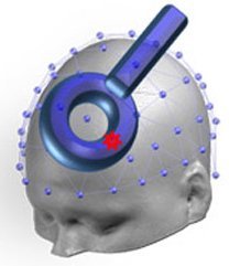
Schematic representation of a typical round TMS coil placed over the left dorso-lateral prefrontal cortex.
Example of publication
Spierer L, Manuel AL, Bueti D, Murray MM.
Contributions of pitch and bandwidth to sound-induced enhancement of visual cortex excitability in humans.
Cortex. 2013, 49(10):2728-34 [Pubmed] -
Transcranial direct current stimulation
Transcranial direct current stimulation (tDCS) is a non-invasive brain stimulation method allowing to modulate cortical excitability with a weak constant current delivered with two electrodes placed on the scalp. Specific configurations of the anodic or cathodic electrode enable one to either increase or decrease the excitability of some portion of the cortex. As with transcranial magnetic stimulation, the tDCS help establishing whether a given brain area is necessary for a given sensori-cognitive process.
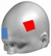
Schematic representation of a typical tDCS montage with the anode (red, positively charged electrode) placed over the left dorso-lateral prefrontal cortex and the cathode (blue, negatively charged electrode) over right supra-orbital areas.